

The Daily Free Press recounts the HUBWeek event in which Center Director Bruce Rosen and medical illustrator Danny Quirk spoke about the intersectionality of human anatomy and visual art.
Optical imaging could lead to fewer unnecessary biopsies in breast cancer screening
February 9, 2015
 |
|
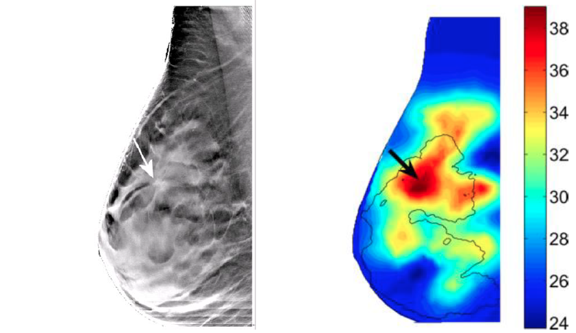
An optical imaging technique developed in the MGH Martinos Center could help to reduce the number of unnecesary biopsies in breast cancer screening. As shown on the right, the technique reveals the hemoglobin concentrations associated with the formation and growth of tumors. A scan with the X-ray-based technique digital breast tomosynthesis is shown on the left. The arrow in each image points to the 2-cm invasive carcinoma in the breast. |
The prevalence of unnecessary biopsies is an ongoing problem in breast cancer screening. Some three quarters of biopsies on lesions found in the breast turn out to be negative, resulting in undue stress for patients – stress in undergoing the procedures themselves and then in having to wait to hear the results – while contributing to higher overall healthcare costs.
Now, an emerging technology is showing promise in helping to reduce the number of unnecessary biopsies. The technology, called tomographic optical breast imaging, or TOBI, can measure variations in blood supply and metabolism associated with the formation and growth of tumors. This information can be used to distinguish tumors from noncancerous lesions, and thus potentially rule out biopsies on those lesions.
“TOBI finds cancers in a new dimension – tissue function – and therefore nicely complements conventional structure-based imaging modalities,” said Qianqian Fang, an Assistant Professor in Radiology at Harvard Medical School and one of the principal investigators leading the optical breast imaging effort in the Athinoula A. Martinos Center for Biomedical Imaging at Massachusetts General Hospital. “It is noninvasive, safe and potentially low-cost, and it does not use ionizing radiation, all of which makes it a good candidate for wide utility in cancer screening and related applications.”
Fang and other researchers in the Optics Division of the Martinos Center have been working with colleagues in the Breast Imaging Division in the MGH Department of Radiology to test the method in conjunction with existing X-ray technology. While a standalone TOBI system demonstrated sensitivity to breast cancer, the investigators knew they could boost the sensitivity even further by incorporating structural information about the breast, particularly from an advanced X-ray mammography technique developed by the hospital’s Dr. Daniel Kopans: 3D digital breast tomosynthesis, or DBT.
This combination of imaging modalities – known in imaging circles as the ‘multimodal’ approach – has precedent in the clinically widely adopted PET-CT (positron emission tomography-computed tomography). Like PET-CT, the new breast imaging method integrates both structural and functional information. Optical imaging provides the latter, including information about the metabolic demands of tumors as well as the increased demand for blood supply. The X-ray-based technique offers structural guidance, helping to localize the tumor while facilitating increases in optical contrast and resolution.
Importantly, an imaging session with the integrated system does not take any longer than a conventional X-ray scan, so it does not involve any additional discomfort for the patient. The ‘optodes’ – the light sources and detectors used for optical imaging – easily slip onto the mammogram paddle used for X-ray imaging while recent advances in TOBI technology enable simultaneous acquisition with the two modalities.
 |
|
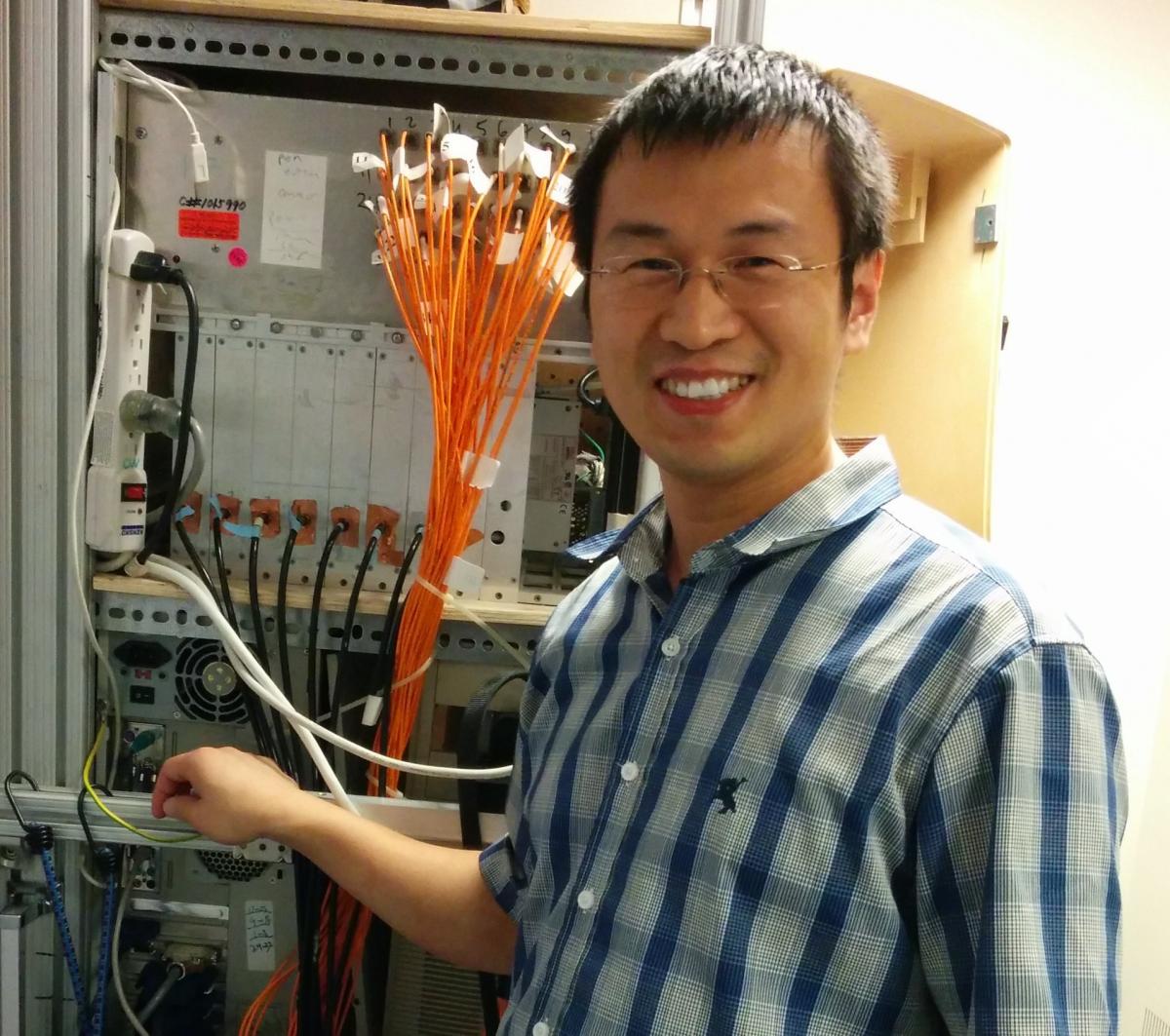
Qianqian Fang, one of the principal investigators in the Optics Division of the MGH Martinos Center, helped to develop the optical imaging technology. |
To test the technology, the researchers have performed scans in 450 patients, more than 200 of them with tumors. “We’re now in the process of analyzing this data set,” Fang said, “trying to identify the sensitivity and specificity of this combined approach and compare it with the performance of X-ray mammography alone.”
They don’t yet have a final specificity, he said, but they are seeing significant differences between lesion types in the various optical properties they are measuring. This will of course aid in distinguishing between those lesion types.
The investigators will report their latest progress with the combined imaging system in a presentation this week at SPIE Photonics West, the premier biomedical optics conference, held in San Francisco.
“We will describe the multimodal imaging protocol we’re using and summarize our efforts to establish an optimal compression procedure, with the highest sensitivity to the presence of breast lesions,” said Bin Deng, the postdoctoral fellow in the Martinos Center’s Optics Division who has conducted much of the recent research and will give the presentation.
Once they have established this, they hope to move on to clinical trials with the technology: for diagnosis and later for monitoring of a tumor’s response to therapy.
The latter is an exciting potential application of the multimodal imaging method. “We have shown with the optical-DBT imager that, during breast compression, malignant tumors behave distinctly differently than the surrounding normal tissue, and that these differences fade in tumors responding to chemotherapy,” said Stefan Carp, an Assistant Professor of Radiology at Harvard Medical School and another of the principal investigators involved in the optical breast imaging effort at the Martinos Center.
“Thus the technology can reveal whether a therapy is working – even during the course of the therapy," he said. "This can help oncologists personalize treatments by switching to alternate therapeutic agents or discontinuing ineffective ones, limiting unnecessary side effects and toxicity.”