

The Daily Free Press recounts the HUBWeek event in which Center Director Bruce Rosen and medical illustrator Danny Quirk spoke about the intersectionality of human anatomy and visual art.
New Optical Imaging Tool Could Allow Neuromonitoring Of Stroke And Other Patients At Bedside
September 6, 2016
 |
|
Members of the Optics Division in the MGH Martinos Center described the technology in a recent paper. |
The optical imaging technique diffuse correlation spectroscopy (DCS) has shown tremendous promise for neuromonitoring in infants and children—offering a means to measure cerebral blood flow in, for example, the neonatal intensive care unit. But translating it for use in adults has presented a much greater challenge.
In a paper published last week in the journal Optica, a team of researchers at the MGH Martinos Center described an advance that, for the first time, has made this possible.
Like its sister technique, near-infrared spectroscopy (NIRS), DCS suffers limitations when operating with a continuous-wave laser. Namely: It can’t provide the information needed to separate out the signal from the skull and the scalp, which can contaminate the signal you actually want to see. (Continuous-wave is less problematic in infants and children because their skulls are generally thinner; the extracerebral contribution is larger in adults and, for this reason, the sensitivity to the brain is reduced.)
As with NIRS, using either frequency-domain or time-domain techniques—where you can extract more information from the signal by varying the amplitude or duration of the laser—could help to overcome these limitations. But for assorted, often esoteric reasons, this is much more difficult with DCS than it is with NIRS.
“Nobody believed you could do it,” said Maria Angela Franceschini, an investigator in the Optics Division of the MGH Martinos Center for Biomedical Imaging, an associate professor of radiology at Harvard Medical School, and the senior author of the Optica paper. “We decided to try because there was a chance we could.”
It turned out they were right. In the study, Franceschini and colleagues describe an elegant solution to a seemingly intractable problem with DCS in the time domain: the limited coherence that results from the use of a pulsed laser.
They were able to address this by reimagining their approach and incorporating ideas from other techniques. But even after they sorted out the conceptual issues, they had practical matters to attend to. One example: The laser they needed didn’t exist; because there was no real demand for a laser particularly suited to DCS in the time domain, manufacturers hadn’t yet made one. Undeterred by this, Jason Sutin, a graduate student in the Center, built the laser himself. Similarly, the detectors typically used with DCS weren’t stable enough to work in the time domain, so the investigators employed specialized detectors developed by a lab in Milan, Italy.
One of the advantages of the instrument they ultimately described is that users can perform both time-domain DCS and time-domain NIRS using the same laser source and the same detectors—“so from a single measurement you can obtain both tissue optical properties and blood flow,” Franceschini said.
The ability to measure these two parameters, simultaneously and directly in the brain, could yield a number of benefits—especially in the clinic, where doctors need to know when changes in cerebral blood flow are putting patients at risk.
Among the possible applications: monitoring of the brain at bedside in stroke or trauma patients or other patients in neurointensive care. Also, surgeons could use the technology in the operating room, to track responses to anesthesia or to keep an eye on perfusion during cardiac procedures that involve bypass.
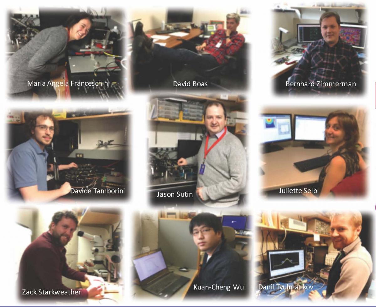
Authors of the Optica paper include the Martinos Center’s Bernhard Zimmerman, Danil Tyulmankov, Davide Tamborini, Kuan Cheng (Tony) Wu, Juliette Selb and David Boas, as well as Franceschini and Sutin.