| Home | People | Publications | Research | Resources |
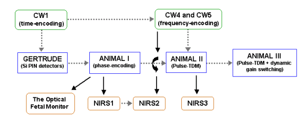
The Optical Fetal Monitor
The Optical Fetal Monitor (OFM) was developed in an attempt to quantify fetal blood oxygenation noninvasively, through a simple transabdominal measurement. It measures the amount of light absorbed by the fetus at two different near-infrared wavelengths. The fetal modulation signal can then be uniquely resolved from the maternal signal by its frequency content.
The optical flux reaching each detector optode will be modulated by both maternal and fetal cardiac activity as the elastic arterial walls expand slightly and then relax following each ventricular contraction. This appears as a real-time modulation of blood volume within the tissue. Any light passing through tissue perfused by these vessels will exit with a small amplitude modulation in the shape of an inverted sawtooth. A sharp drop in optical signal during ventricular contraction (systole) is followed by a slower signal rise as the blood passively exits the capillaries and drains into the venous compartment on its way back to the heart (diastole).
Since the mother and fetus have separate hearts and thus distinct heart rates, the blood passing through the fetus will be “frequency tagged” by the fetal heart rate. A Fourier transform can then be performed on the detected signals to discriminate the fetal signal from the maternal signal, since the mother’s heart rate, and hence her contribution to the amplitude modulation, should be far slower. By performing measurements at two or more optical wavelengths, it should be possible to measure the oxygen saturation of both the maternal and fetal blood over time using standard pulse oxygenation calculations. Although absolute calibration is probably not possible due to optical shunting errors, a decrease in fetal oxygenation, which may indicate inadequate placental or fetal perfusion, should be detectable.
The prototype system consisted of a single wavelength light source (an 808nm fiber-coupled laser diode) and four separate optical detectors (Burr-Brown OPT209). Synchronous demodulation was used to both reduce extraneous interference from ambient light sources and to improve the signal to noise ratio of our measurements. The optical source was electrically modulated at a frequency of around 2kHz or so. This frequency could be varied slightly to avoid aliasing with harmonics from AC-powered light sources. The detectors simply detect any in-band optical flux they see, which can include sunlight, electric lighting, or even the light from computer CRT displays. Somewhere amongst all of that lies our weak modulated optical signal.
We retrieve that signal by first passing the detector output through a high pass filter (to remove any slowly varying components produced by sunlight or incandescent lamps) and then we feed the output into a synchronous detector. This circuit is similar to an RF mixer: it electrically multiplies the filtered detector signal with a “carrier” signal using analog switches. This carrier signal is the same 2kHz signal used to gate the source on and off. The only difference is that a slight time delay is added to the carrier to make up for the time it takes for the optical signals to propagate through the detector circuitry. This way the gated light from the optical source and the carrier signal arrive at the synchronous detector at exactly the same time. This is important because the synchronous detector will act as a rectifier for the gated optical signal – and only that signal. All other stray light signals (which are all uncorrelated to the gated source) will exit the synchronous detector as frequency-shifted AC signals. But the small signal component from the gated source (the same one which hopefully passed through both the mother and the fetus) will be “synchronously” rectified, and will appear as a small DC voltage.
Since this synchronous process is linear, the DC level tracks the amount of gated light reaching the detector (yes, the demodulator multiplies two signals, but think of it as multiplying the gated source signal by the number “1”). The output of the synchronous detector then passes through a lowpass filter to eliminate all the other frequency-shifted AC signals which we don’t want.
The bandwidth of this filter is important, since it sets the frequency response of our measurement and it thus determines the noise floor. A wide bandwidth would allow us to measure lots of heartbeat harmonics, but at the price of lower sensitivity due to a higher noise floor. A narrow bandwidth would yield much better sensitivity, but might not be fast enough to catch the second or third heartbeat harmonics, which are important in discriminating the fetal signal from the maternal signal.
A drawing of the prototype OFM unit is shown below. All functions are controlled by the computer. The 808nm laser source module generates the optical signal which reaches the optode assembly through a single 1mm diameter silica fiber. Its output is modulated by an oscillator located in the control module. The four detectors, each spaced one inch (25mm) apart on the optode assembly, couple to the patient through short lengths of 3mm diameter PMMA optical waveguide. The detector outputs travel back through the umbilical cable to the control module, where each output goes to its own amplifier. Since the detector located farthest from the source will receive the least light, its signal receives the most gain (1000x). The more proximal detectors receive proportionally less gain (100x, 10x, 1x). This is necessary, since the amount of signal reaching the distal detector can be ten thousand times weaker than that reaching the most proximal one.
Scaling the gain is an easy way of conserving our dynamic range for more important things, like fetal heartbeat signals. Since each patient is likely to have a unique distribution of adipose tissue and an unknown vascular structure, the actual detector signal levels will vary from mother to mother, and our dynamic range had to allow for this.
The four synchronously rectified and filtered DC outputs then pass from the control module to the signal processing computer. They are digitized at a constant rate of 1000Hz and stored in memory. While this is going on, the computer grabs a portion of the data and uses that time sequence to generate a Fourier transform, which is then displayed. This presents the data, sampled in the time-domain, as a 2-D plot in the frequency-domain. Signal power components modulated at different frequencies now appear distinct from each other. In this way we can measure the modulation created by both the mother’s heartbeat and the fetal heartbeat as discrete quantities – which is important for blood oxygenation measurements.

A diagram of the Optical Fetal Monitor, showing the modular format. It consists of five modules: the laser source module, the control module, the optode assembly, the signal processing computer, and the system power supply. Optical power from the laser source module is fed to the optode assembly in contact with the mother’s abdomen. The weak optical signals reflected and scattered back to the skin surface (curved red lines) are detected by four OPT209 detectors, which amplify the photodiode current and buffer the resulting signal. These four signals then travel through shielded cables to the control module, where they are synchronously rectified and then filtered. The filtered signals are then sampled by a PCMCIA ADC card in the computer and stored in memory for later analysis. Approximately 98% of the light traveling from the source to the most distal detector will pass through the fetus en-route.
The following paper reports on a study using the Optical Fetal Monitor.

(return to the continuous-wave DOT instrument home page)